Types of Skin Cancer
Considering how prevalent skin cancer is, it is exceedingly important to pay attention to your skin and visit a dermatology-trained provider if anything looks abnormal. That being said, it can be difficult to self-determine if a mole is just a mole—or if something more serious is at play.
Read on to learn more about types of skin cancer and how to recognize when it’s time to schedule an appointment.
The ABCDEs of skin cancer
Because most of us have freckles, skin discolorations, moles, and birth marks somewhere on our bodies, it’s important to find a balance between not being an alarmist about our skin and being properly educated so we know when we should get checked out.
The ABCDE checklist has been shown to be an incredibly helpful tool in assessing your own skin and detecting melanoma, specifically. If you note any of these five signs, give your doctor a call and ask for an evaluation.
- Asymmetry: Do both sides of the mole look the same? Or are they mismatched?
- Border irregularity: Are the edges of your mole uneven, notched, or distorted? Do they feel raised or scalloped?
- Color variation: Has the color of your mole changed or does it have different colors throughout? Do you see red, blue, white, or black?
- Diameter of greater than 6mm (about 1/4 inch): Is your mole larger than a pencil eraser?
- Evolution: Did your mole change in any way? Is the size, color, or elevation different? Are there new symptoms, like bleeding?
Recognizing cancerous lesions
Benign lesions & precancerous tumors
Not all skin lesions are cancerous or at all problematic. In layman’s terms, these benign lesions are the moles, “beauty marks,” or other skin markings that we commonly see that have a normal color, size, and border that remains consistent.
The most common lesions are benign junctional, compound, and intradermal nevi. Visually, they can mimic the appearance of melanoma, seborrheic keratosis, and solar lentigines. If there is any question about the malignancy of the lesion or you would prefer to have it removed, your provider may recommend a biopsy or excision procedure.
Pre-cancerous lesions can lead to melanoma and include atypical and dysplastic nevi. Actinic keratosis may become a squamous cell cancer, while trichoepithelioma can mimic basal cell carcinoma and keratoacanthoma resembles squamous cell carcinoma. We often recommend having these common lesions treated or removed.
Basal cell carcinoma
Basal cell carcinoma is the most common type of malignant lesion. When diagnosed and treated early, it can typically be completely removed before the cancer spreads. While basal cell cancer is not typically a fast-spreading cancer, it can run fairly deep beneath the skin and lead to additional issues if left untreated.
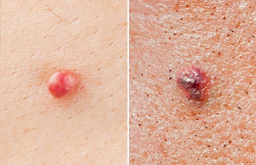
Basal cell carcinoma occurs in the deepest layers of the epidermis in basal cells. At the surface level, lesions appear as shiny bumps, patches of red, or an inflamed sore.
Squamous cell carcinoma
Squamous cell carcinoma is a more invasive cancer than basal cell carcinoma, though it has a similarly high cure rate if caught early. Most common in those who have experienced many years of sun exposure, squamous cell carcinoma is the second most common form of skin cancer with over a million diagnosed cases per year in the United States.
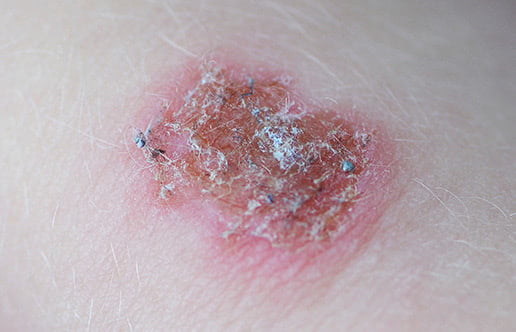
Formed in the squamous cells in the epidermis, this type of skin cancer usually appears as a sore, scabby patch of skin, wart, or elevated area prone to bleeding on contact.
Melanoma
Melanoma is the most dangerous, aggressive form of skin cancer. Again, detecting and treating melanoma early is key to a good prognosis, as it can spread very quickly. If left untreated and allowed to spread more deeply into other tissues, melanoma can be fatal.
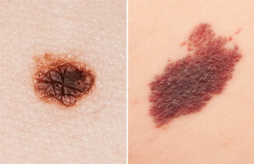
For some patients, the first sign of melanoma is a change in an existing mole—it’s bigger, has changed colors, or feels differently than before. However, melanomas can appear in the form of a new lesion, which is why it’s so important to reach out to your dermatology-trained provider if you notice anything new.
How to perform a skin cancer self-exam
Examining your skin head-to-toe around once a month is one of the best ways to detect skin cancer early and increase the likelihood of successful treatment—and it’s easy!
In a well-lit room, start by examining your face, neck, and chest, particularly areas around the ears, nose, and mouth. Areas of your skin that are most exposed to the sun on a daily basis have a greater risk of skin cancer, so be sure to pay particular attention to these spots. Move down each arm and hand, bending and angling your arm so you can view the elbows and forearms. Don’t skip over your armpits and between the fingers!
Take the same care as you look over your torso, upper thighs, and legs, taking care to check in natural skin folds, such as under the breasts and around the groin. Even though these areas may not see the sun, it’s important not to overlook them. Move down to your feet, top and bottom, and between your toes.
Enlist the help of a loved one or a full-length mirror to repeat the full exam on the backside of your body. Don’t skip over your scalp! Using a hairdryer, throughly examine your scalp visually and through touch. If at any point you notice any new growths or changes in existing moles, document the changes for your physician.
What to do if you suspect skin cancer
If you’ve noticed anything of concern on your skin, don’t hesitate to contact your dermatology-trained provider and set up an appointment. As one of our dermatology services here at Berks Plastic Surgery, our Dermatology Physician Assistant can evaluate and diagnose skin cancer and perform a biopsy to test for cancer cells and remove cancerous lesions as needed.